Fig. 1
From: Peripheral neuropathy in a case with CADASIL: a case report
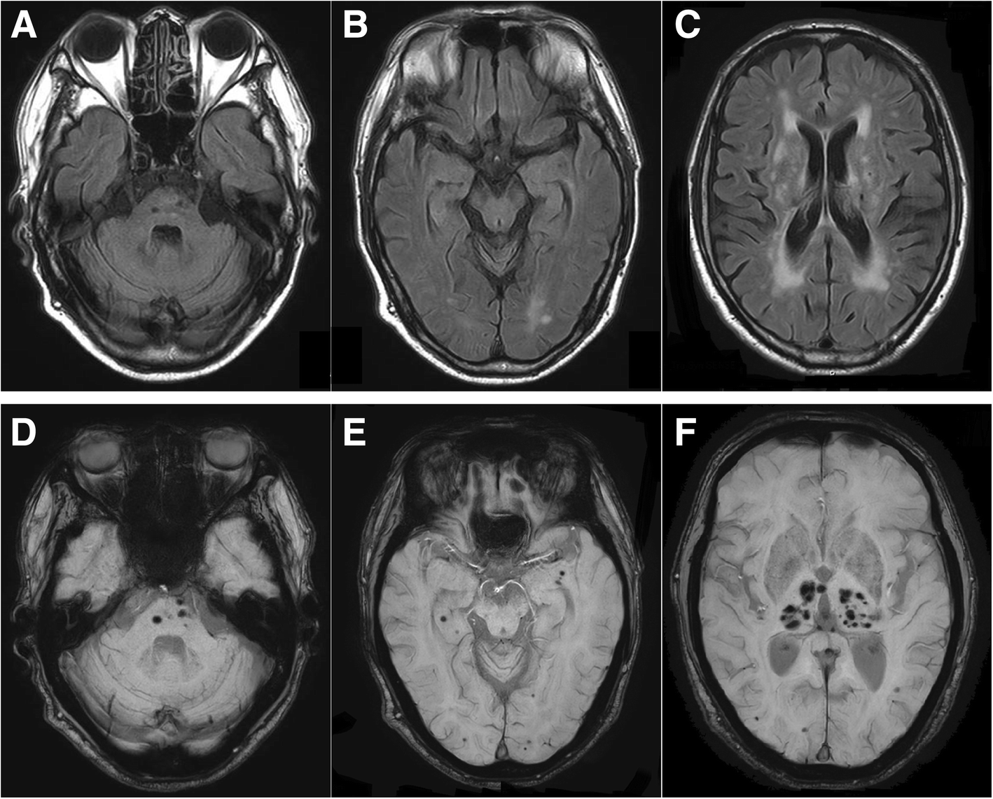
Brain-MRI findings of our patient. a–c Axial FLAIR image showing the bilateral ischemic lesions in pons, periventricular white matter and deep white matter. d–f: Axial susceptibility-weighted imaging (SWI) showing multiple micro-bleeding lesions in pons, bilateral subcortical white matter and bilateral thalamus